Exploring Brain Functions and Their Connections


Intro
The human brain, often described as the pinnacle of evolution, is an organ of extraordinary complexity. It's not mere gray matter confined in a skull; rather, it orchestrates thoughts, emotions, memories, and movements, all intertwined in an elaborate web of neural connectivity. Understanding the brain's mapping serves as a foundational step toward comprehending how we define our experiences and interactions. From the rhythmic firing of neurons during a stimulating conversation to the quick recall of a long-lost melody, the interconnections of brain functions play a pivotal role in every aspect of our lives.
Research Overview
Key Findings
Recent advancements in neuroscience have unveiled startling insights into the brain map. Studies have shown, for instance, how specific areas are responsible for distinct cognitive functions, yet these regions seldom operate in isolation. The brain thrives on collaboration; a single thought could engage multiple areas working in unison. This interconnectedness reveals how imbalances can lead to disorders, emphasizing the relevance of understanding these dynamics. For instance,
- The prefrontal cortex is crucial for decision-making, yet it draws on emotional contexts processed by the amygdala.
- Spatial orientation, managed by the hippocampus, can also affect how we interact with our environment.
This multi-dimensional approach not only enhances our knowledge but also lays the groundwork for developing targeted treatments in neurology and psychology.
Study Methodology
The methodologies employed in brain mapping have evolved drastically over the years. Techniques such as functional magnetic resonance imaging (fMRI) and positron emission tomography (PET) allow researchers to visualize brain activity during various tasks. These technologies capture real-time changes in blood flow and metabolic activity, painting a dynamic picture of the brain in action.
Furthermore, advancements in electrophysiological techniques provide detailed measurements of electrical activity in specific brain regions. This combination of imaging and electrical monitoring has led to significant breakthroughs in understanding both normal brain function and the pathological underpinnings of mental health disorders.
"Understanding the brain is not just a question of anatomy; it is a process of mapping connections and functions that define who we are as individuals."
Background and Context
Historical Background
The journey of studying the brain's mapping extends back to ancient civilizations, where philosophers like Hippocrates posited that the brain was the seat of consciousness. However, it wasn't until the 19th century that scientific inquiry began to take shape. Pioneers such as Paul Broca and Carl Wernicke identified key areas tied to speech and language, establishing a foundation for modern neuroanatomy.
Current Trends in the Field
Today, research in brain mapping has reached new heights with the advent of artificial intelligence and machine learning. These technologies facilitate the analysis of vast amounts of data, leading to finer distinctions between regions once viewed as homogenous. As we continue to peel back the layers of the brain's intricacies, emerging trends are revealing the interplay between genes, environment, and neural activity in shaping behavior and cognition.
Infinity loops of inquiry are now being forged, as the lines between disciplines blur and novel collaborations enhance our understanding of the human experience. Thus, the exploration of brain mapping not only illuminates the path to unraveling the complexities of mental health conditions but also holds remarkable promise in advancing therapeutic techniques.
Prologue to Brain Mapping
The brain, a complex organ, is often likened to a master conductor orchestrating the symphony of human experience. Understanding brain mapping is crucial in deciphering this intricate work of art, as it provides insights into how various regions interconnect and collaborate to produce behaviors, thoughts, and emotions. Through brain mapping, we peel back the layers of mystery surrounding cognitive functions, revealing the physical and chemical foundations of human consciousness.
In this article, we aim to explore the vital aspects of brain mapping, focusing on its definition, historical context, and why it remains a cornerstone in neuroscience. This exploration isn’t just for the academics or medical professionals; it also has implications for anyone interested in the nature of the human experience—the very fabric of who we are.
Definition of Brain Mapping
Brain mapping can be defined as the systematic way of representing the various anatomical and functional aspects of the brain. It helps identify which parts of the brain are responsible for specific functions, such as memory, emotion, and sensory processing. This practice incorporates a variety of techniques like neuroimaging and electrophysiology to illustrate how neural pathways interconnect and interact.
For instance, scientists use functional magnetic resonance imaging (fMRI) to analyze brain activity while a subject engages in different tasks. By focusing on blood flow changes, researchers can visualize which areas of the brain are being utilized during specific activities, creating a useful map of brain functions.
Historical Context and Evolution
The journey of brain mapping has evolved significantly since the days of early anatomical research. Historically, observations of the human brain began with figures like Hippocrates and Galen, who postulated roles for brain structures based on surface examinations and bodily functions. Fast forward to the 19th century, when pioneers such as Paul Broca discovered localized functions in the brain, particularly in speech production, laying groundwork for modern cerebral localization studies.
As technology advanced, so did our understanding. The invention of the electroencephalogram (EEG) in the 1920s allowed for the recording of electrical activity in the brain, opening new avenues in brain mapping. Later, the advent of imaging techniques like PET scans and MRI further revolutionized the field, enabling researchers to visualize brain structure and function without invasive procedures.
Today, brain mapping is a collaborative field that integrates neurobiology, cognitive science, psychology, and advanced imaging techniques. Each stride made in this domain leads us closer to understanding complex neurological disorders and enhancing clinical practices, underscoring the importance of comprehensively grasping how our brain orchestrates its functions.
Anatomy of the Brain
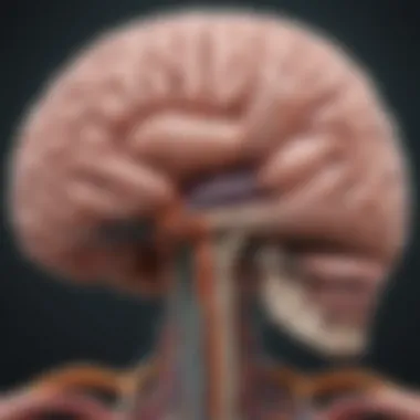
Understanding the anatomy of the brain is like having a map in a foreign land. It reveals the diverse territories, the major landmarks, and the intricate connections most of us never realize until we dig deeper. Each region plays a significant role in not just individual functioning, but in how we collectively interpret and engage with the world. By exploring the anatomy of the brain, we can develop a clearer comprehension of how its various parts work together in concert—shaping our behaviors, emotions, and cognitive processes. This is particularly essential in the context of medical science, where mapping brain anatomy can guide treatment plans for neurological disorders, and deepen our understanding of human psychology.
Major Regions of the Brain
Cerebrum
The cerebrum stands tall as the largest and most prominent part of the brain. It is where the magic happens—the realm of thought, sensation, and voluntary movement. Covering its surface is the cerebral cortex, a folded layer that’s akin to a crumpled paper. The mechanisms within, both electric and chemical, work tirelessly to process an astonishing amount of information. Its key characteristic lies in its functional specialization; it houses various lobes, each responsible for different tasks. For instance, the frontal lobe tackles executive functions while the occipital lobe is primarily about vision.
A unique feature of the cerebrum is neuroplasticity, which denotes its ability to reorganize itself in response to learning or injury. This characteristic tells us why rehabilitation is so vital after brain injuries. Such flexibility can be viewed as an advantage, providing hope and a pathway for recovery. However, the underlying complexities of cognitive processes can also pose challenges in understanding the full extent of brain damage.
Cerebellum
Often referred to as the "little brain," the cerebellum is tucked beneath the cerebrum, yet plays a pivotal role in fine-tuning motor activities. Its primary function involves coordinating voluntary movements, balance, and posture. The cerebellum is key for fluidity—think of it as the director of a graceful performance where each movement flows seamlessly into the next.


Its key characteristic is its structure; compared to the cerebrum, it has more neurons packed within a smaller space. This unique feature allows for precise control over muscle memory and balance. The implications of a dysfunctional cerebellum are significant, as issues here can lead to staggering, clumsiness, or lack of coordination. So, in this article, we highlight the cerebellum to emphasize not only its contributions to motor function but also how dysfunction can impact everyday life.
Brainstem
The brainstem may not be as glamorous as the cerebrum or cerebellum, but it holds the reins of survival. It serves as the gateway between the brain and the spinal cord, controlling many automatic functions that are essential for life—such as breathing, heartbeat, and blood pressure. A remarkable feature of the brainstem is its role in regulating consciousness and alertness, which is critical when considering sleep cycles or consciousness-related disorders.
Its intricate design allows it to relay signals between the brain and the rest of the body, integrating sensory information and motor control. While it may not be the center of intellectual activity, damage to the brainstem can have fatal consequences, underlining its importance in both basic body functions and overall neuronal health.
Understanding Gray and White Matter
Gray matter contains most of the brain's neuronal cell bodies and is involved primarily in muscle control and sensory perception. White matter, on the other hand, is composed of myelinated axons that facilitate communication among different brain regions. The contrast of these two types of matter is not just structural but functional. Understanding their roles provides insight into conditions such as multiple sclerosis, where white matter damage disrupts communication pathways. By discerning the functions associated with gray and white matter, we gain a comprehensive understanding of the brain's functional physiology.
Cognitive Functions and Brain Areas
Understanding cognitive functions is key to grasping how our brains work. Each area of the brain manages different tasks, and recognizing these can lead to breakthroughs in various sectors like education, psychology, and even artificial intelligence. When examining cognitive functions, one sees the interplay of emotions, decision-making, and memory processes. This understanding leads to better educational practices and more effective mental health therapies, hence enriching human experience on multiple fronts.
The Role of the Frontal Lobe
The frontal lobe is often considered the brain's command center. It orchestrates many higher-level functions that separate us from other species.
Executive Functions
Executive functions encompass a range of cognitive abilities including organization, planning, and prioritization. High-functioning executive skills are what allow individuals to put thoughts into action smoothly. This is essential for setting goals and achieving them. A notable aspect of executive functions is self-regulation. It helps maintain focus and control impulsiveness, a characteristic that’s vital in educational realms. Its profound impact in clinical assessments enables tailored interventions. Awareness of one’s executive prowess can make a world of difference—assisting professionals to develop strategies that can lead to improved outcomes in their fields.
Decision Making
Decision-making hinges considerably on the frontal lobe. One aspect of this is the ability to weigh options and forecast potential outcomes. Often, risk assessment during decision-making showcases the brain’s capacity to evaluate consequences, which is essential in personal and professional domains alike. This characteristic plays a vital role in leadership and management roles. However, despite its strengths, decision-making can be influenced by emotional states, which might muddle the logical process. Thus, acknowledging factors that guide decision-making can raise drama in everyday engagements, enriching our dialogues on cognitive paths.
Emotional Control
Emotional control is another significant aspect of the frontal lobe’s functions, relating to how we manage our reactions and interactions. It helps discern which emotions to express or repress during various situations. One crucial element of emotional control is empathy, which plays an impactful role in how we connect with others. Although this function can enhance interpersonal relationships, it does have its drawbacks. Excessive emotional regulation may lead to the suppression of feelings, potentially causing inner turmoil. Understanding this balance provides insights into emotional intelligence—especially relevant in therapy and counseling practices.
Language Processing in the Brain
Language processing is a fascinating domain as it intertwines cognitive functions with communication abilities.
Broca's Area
Broca's Area, found in the frontal lobe, is vital for speech production. This aspect facilitates the translation of thoughts into coherent language. A key characteristic of Broca's Area is its role in grammatical structuring, which is crucial for effective verbal exchanges. Its importance in this article cannot be overstated as communication is foundational in all human relations. One unique feature of Broca's Area is its vulnerability to damage, which can lead to speech impairments known as aphasia. Such conditions highlight the critical link between brain mapping and rehabilitation strategies in language recovery.
Wernicke's Area
Wernicke's Area, located in the temporal lobe, complements Broca's Area by processing and understanding spoken language. It allows individuals to follow conversations effectively and comprehend verbal cues. The distinctive attribute of Wernicke's Area is semantic processing, integral in ensuring that words carry the intended meaning. Its consideration here is beneficial as it sheds light on patterns of language disorders, enhancing therapeutic approaches for patients with communication challenges. However, deficits can lead to fluent aphasia, a condition where individuals produce grammatically correct speech but fail to convey the desired message—a fascinating paradox of language development.
Memory and Learning Mechanisms
Memory is where learning springs forth, enabling humans to navigate the complexities of daily life.
Hippocampus Involvement
The hippocampus is crucial for memory formation and storage, particularly for long-term memories. Much of its work revolves around consolidating new information with existing knowledge bases. A notable aspect of the hippocampus is spatial memory, which aids in navigation and environmental understanding. It's a pivotal component of this article, as it illustrates connections between cognition and daily experiences. However, its fragility means damage can lead to difficulties in forming new memories, raising essential questions related to dementia and Alzheimer’s.
Short-term vs Long-term Memory
Short-term and long-term memory intertwine seamlessly to create a holistic memory system. While short-term memory allows for immediate retention of information, long-term memory stores knowledge for extended periods. The process that distinguishes these two types is encoding, which is crucial for effective learning. Understanding this difference is both beneficial and relevant, particularly in educational settings where strategies can be tailored accordingly. However, challenges arise in retention of long-term memories, often causing frustration, depending on various cognitive and environmental factors.
"Understanding the consequences of brain functionality expands the horizons of cognitive therapies and educational methodologies, bridging theory with real-world applications."
In summary, delving into cognitive functions and their corresponding brain areas illuminates complex pathways of human behavior and interaction. Recognizing the specific roles of each area allows for more effective teaching, better therapeutic practices, and a deeper appreciation of human cognition.
Sensory Functions and Their Mapping
Understanding sensory functions and their mapping is a critical piece of the overarching puzzle that is brain function. Sensory processing is what allows us to interpret and interact with our environment, and mapping these functions provides insight into not only how we perceive the world but also how those perceptions influence our behaviors and reactions. This section will dissect the key components of sensory processing, including how diverse brain areas contribute to a holistic understanding of sensory information.
- The mapping of sensory functions is essential for diagnosing and treating conditions like sensory processing disorders.
- It serves as groundwork for developing assistive technologies that can enhance or replicate sensory experiences for affected individuals.
- Beyond clinical applications, insights gained from sensory mapping enrich our understanding of human perception and cognition, linking our sensory experiences to memory, emotion, and decision-making.
Mapping sensory functions can reveal how interconnected our experiences are across different sensory modalities, illuminating the brain’s intricate web of processing activities.
Visual Processing Centers


Visual processing is a complex function involving multiple brain regions, each playing a unique role in the famed journey of light becoming perception. The journey begins when light hits the retina and is transformed into electrical signals. These signals travel along the optic nerve and relay crucial information to the primary visual cortex, located in the occipital lobe. It’s here, in the early stages of processing, that simple features of visual stimuli, such as lines and colors, are interpreted.
As we delve deeper, we see that aspects of visual processing extend beyond mere recognition of shapes and colors. Secondary visual areas further refine this data, enabling higher-level functions like motion detection, object recognition, and even depth perception. One area of particular interest is the parahippocampal gyrus, which is integral in recognizing places and spatial awareness. This isn’t just academic; impairment in these visual centers can lead to conditions like visual agnosia, where individuals can see but fail to recognize objects.
Auditory Processing: The Temporal Lobe's Role
The auditory processing centers, predominantly situated in the temporal lobe, serve as the brain's mechanism for interpreting sound. Much like visual stimuli, sound waves are converted into electrical signals that travel through the auditory pathway. The primary auditory cortex decodes these signals, allowing for the detection of pitch and volume. However, auditory processing doesn’t stop there.
Secondary regions within the temporal lobe, such as Wernicke's area, are crucial for processing the meanings of sounds, enabling us to understand speech and other complex auditory signals. The intricate connection between auditory perception and language comprehension highlights just how intertwined these functional pathways are. Moreover, studies indicate that damage to specific portions of the auditory cortex can lead to difficulties in distinguishing between similar sounds, a phenomenon affecting many musicians and language learners.
The Somatosensory Cortex
The somatosensory cortex, nestled just behind the frontal lobe, serves as the brain's hub for processing touch and proprioceptive information. This area is responsible for interpreting signals from the skin, muscles, and joints, which helps in understanding our body's position and movement. The somatosensory system is not only about feeling; it’s about processing the very nature of the stimuli that our bodies encounter.
Different parts of the somatosensory cortex are correspondingly mapped to different body areas, creating a phenomenon often referred to as the 'homunculus'. This mapping illustrates just how much brain real estate is devoted to our sensory experiences. For instance, our lips and fingertips occupy a much larger representation than do, say, our elbows or knees, highlighting their importance in our interactions with the environment.
The significance of the somatosensory cortex extends to clinical practice as well. Disorders such as chronic pain syndromes can be traced back to the malfunctioning of this sensory map, complicating diagnosis and treatment plans. Understanding the layout and functions of the somatosensory cortex thus not only informs the scientific community but also has real-world implications for patient care.
Motor Functions and Brain Coordination
Motor function, often underappreciated, plays a pivotal role in how humans interact with their environment. Understanding motor functions and their coordination provides a crucial insight into not only how we perform tasks but also how various parts of the brain communicate and work together to execute movements. This section will focus on the specific components that drive motor functions, exploring the intricate dance between the brain areas involved, the mechanisms at play, and the implications of this understanding for areas such as rehabilitation and sports science.
In simpler terms, motor functions deal with everything from picking up a cup to performing complex athletic maneuvers. The brain orchestrates these activities by firing signals that prompt our muscles to move, which involves a sophisticated network of regions working in harmony.
The Motor Cortex
The motor cortex is the brain region fundamentally associated with the planning, control, and execution of voluntary movements. It’s located at the back of the frontal lobe and consists of distinct areas that correspond to different body parts. The idea of a homunculus, often illustrated in neuroscience, shows how areas in the motor cortex translate into specific muscle groups. The hand and face occupy a significant amount of this map, highlighting the fine motor skills the brain commands in these regions.
When we talk about the motor cortex, we need to consider its two key parts: the primary motor cortex and the premotor cortex. The primary motor cortex is essential for executing movements, whereas the premotor cortex is more about planning and preparing the brain for movement. Here, complex movements are organized before they are sent to the muscles. For instance, when you decide to throw a ball, your premotor cortex gets busy mapping out the required actions long before you actually release the throw.
Key Functions of the Motor Cortex:
- Motor Planning: Initiating complex sequences of movements.
- Movement Execution: Sending signals to muscles to perform the action.
- Fine Motor Control: Enabling precise movements, particularly in the hands.
Role of the Basal Ganglia and Cerebellum
Beyond the motor cortex, the basal ganglia and cerebellum are critical players in the orchestration of motor functions. The basal ganglia, a group of nuclei located deep within the cerebral hemispheres, are primarily associated with the initiation and regulation of movement. These structures are integral in deciding which movements to execute while inhibiting unwanted ones. When bowling, for instance, the basal ganglia steps in to help you focus only on that swinging motion, preventing your body from doing unnecessary movements.
On the other hand, the cerebellum, often referred to as the "little brain," is crucial for coordination and precision in movement. It’s responsible for functions such as balance and motor learning. After executing a movement, the cerebellum helps to refine that action, making it smoother and more accurate the next time you perform it. A classic example can be seen when learning to ride a bicycle; your cerebellum processes information to correct your balance and coordination dynamically until the skill becomes second nature.
In summary, the interplay between the motor cortex, basal ganglia, and cerebellum exemplifies how our brain coordinates various aspects of movement.
Understanding motor functions is not just about how we move; it's about how our brain and body work together seamlessly, reflecting the intricate networking that allows for complex actions in daily life.
The exploration of motor functions holds significant implications for numerous fields. In rehabilitation, therapies aimed at strengthening connections within these brain regions can lead to improved outcomes after strokes or injuries. In sports science, understanding these pathways enables athletes to optimize their performance through targeted training and practice strategies.
Overall, the integration of motor functions illustrates the brain's capacity to adapt and coordinate. This broadens the horizons for research and a deeper understanding of neurological processes affecting movement.
Methods of Brain Mapping
To fully grasp the intricate workings of the brain, one must dive into the various methods employed in brain mapping. These techniques shed light on how different regions of the brain interact and function. Understanding these methods enables researchers and healthcare professionals to gain deeper insights into brain health, the effects of neurological disorders, and potential treatment methods. This section emphasizes the significance of these mapping methods, highlighting their unique characteristics and contributions to advancing neurological science.
Neuroimaging Techniques
fMRI
Functional Magnetic Resonance Imaging (fMRI) has revolutionized the field of brain mapping. It captures brain activity through changes in blood flow, making it possible to visualize which areas become active during specific tasks. This technique is particularly beneficial as it offers a non-invasive way to monitor brain functions in real time.
A key characteristic of fMRI is its ability to provide high spatial resolution, allowing researchers to pinpoint activity in precise brain regions. Additionally, fMRI is widely employed because it doesn’t involve radiation, making it safer for repeated use.
Among its unique features, fMRI presents a significant advantage in studying cognitive processes, from understanding emotion to tackling complex decision-making tasks. However, it comes with some drawbacks, such as susceptibility to motion artifacts, which can sometimes mislead results if a subject cannot remain still. Still, its ability to reveal the brain's workings during active tasks makes it a cornerstone of modern neuroscience.
PET Scans
Positron Emission Tomography, or PET scans, provides insight into metabolic and biochemical activity in the brain, making it distinct from fMRI. This technique uses a radioactive tracer, allowing researchers to visualize the distribution of metabolic processes in real-time, making it crucial for examining brain function in various states, such as during the onset of disease.
The primary characteristic that sets PET scans apart is their focus on detecting changes in cerebral blood flow—this offers a broader metabolic perspective rather than just localized brain activity. Thus, PET has become a go-to choice when investigating deeper, systemic issues within the brain, especially in cases of degenerative disorders.
One unique feature of PET scans is their capacity to measure neurotransmitter activities, making them especially valuable in research related to mental health. However, the major limitation is the use of radioactive substances, which restricts the frequency of scans for safety reasons. Nonetheless, the depth of information gathered through PET scans is unmatched in many aspects, solidifying its role in brain mapping.


Electrophysiological Mapping
Electrophysiological mapping involves recording electrical activity from neurons. By placing electrodes in or on the brain, researchers can assess neural signaling in real time. This method stands out for its remarkable temporal resolution, making it ideal for studying how quickly and dynamically brain regions communicate.
This technique is particularly useful in understanding the electrical signatures of cognitive functions and how abnormal electrical activity relates to disorders like epilepsy. The detailed data it provides helps map brain regions responsible for specific functions, thus contributing to the development of precise treatment protocols.
Histological Techniques for Brain Mapping
Histological techniques focus on the microscopic examination of brain tissues to understand the brain's cellular structure. This method allows researchers to analyze the composition and connections of various brain areas at a cellular level, providing insights into how different neuron types contribute to brain function.
One popular histological method is staining, which highlights specific types of cells or fibers. This can help clarify the connections between regions and reveal cellular pathology in diseased states. An advantage of histological analysis is its ability to provide a detailed view of the cellular architecture and connectivity that informs the broader understanding of brain functionality.
However, histological techniques often require invasive sampling and can be time-consuming. This limits the immediacy of the insights obtained from such studies. Regardless, they are indispensable in creating a comprehensive map of the brain's structure and its complex network of interconnections.
"Methods of brain mapping not only illuminate the complexities of neural interactions. It also opens doors to therapeutic innovations and better health outcomes."
In summary, the methods of brain mapping underscore an essential aspect of our understanding of neurological functions. From the non-invasive and real-time insights of fMRI to the detailed cellular insights provided by histological methods, each technique contributes uniquely to our growing body of knowledge about the brain's architecture and functionality.
Clinical Implications of Brain Mapping
Brain mapping isn't just an intricate puzzle of regions and connections; it's a significant leap forward in our understanding of the human mind and its disorders. By systematically laying out how different brain areas interact and function, researchers and clinicians can better understand neurological conditions. This ain't just science for the sake of it—there are tangible benefits to society that stem from these studies.
The ability to visualize brain activity helps map out not only normal patterns but also deviations that signal disorders. Such understanding is critical when it comes to diagnoses. The accuracy gained from advanced neuroimaging techniques means that clinicians can identify abnormalities earlier than ever before. For instance, diagnosing conditions like Alzheimer's disease or mild cognitive impairment is becoming more precise, leading to more timely and effective treatments.
Additionally, the implications extend far into the realm of treatment strategies, rehabilitation, and patient education. Understanding the brain significantly influences the development of targeted therapies and establishment of recovery protocols. Ultimately, successful outcomes hinge on how well we interpret and employ the insights gained from mapping the brain.
Understanding Neurological Disorders
Organizations have long been working to uncover the root causes of complex neurological disorders, and brain mapping provides essential tools for this endeavor. Conditions like epilepsy, Parkinson's disease, and multiple sclerosis often show different manifestations depending on the brain areas involved. By creating a map of where these disorders hit hardest, researchers can study their progression and impact on functions.
Take epilepsy, for example. Mapping can help locate the focus of seizure activity within the brain, allowing for more tailored surgical interventions. This precision surgery showcases how crucial brain mapping has become in the realm of treating neurological conditions—surgical outcomes have greatly improved compared to traditional methods. Furthermore, understanding these disorders from a mapping perspective also aids in developing educational resources for patients and families, allowing them to understand the condition better.
Advancements in Treatment Plans
Targeting Specific Brain Regions
Narrowly focused treatments that aim at specific areas of the brain can lead to breakthroughs in managing neurological disorders. When we talk about targeting specific brain regions, it is about precision medicine in the neurosciences. This concept emphasizes more personal treatment strategies, rather than one-size-fits-all approaches. The key characteristic here is its directness: for example, deep brain stimulation can alleviate symptoms in Parkinson's disease by modulating activity in the basal ganglia, enhancing motor function.
However, it's not all sunshine and roses. While this method can be beneficial, it also poses challenges. The unique feature of this approach involves the need for precise placement—misalignment can lead to misinterpretation of results or complications during recovery. Despite these challenges, the advantage it presents in targeting symptoms directly is an exciting prospect for continuing research and application in clinical settings.
Rehabilitation Strategies
Once a disorder has been identified and treated, the next step often involves rehabilitation strategies. These strategies lean heavily on mapping the brain's functioning to aid in recovery. Whether it’s from stroke, traumatic brain injury, or other conditions, rehabilitation seeks to help individuals regain lost skills and independence. The effectiveness of these strategies lies in understanding how brain mapping can inform which areas need the most attention during recovery.
Notably, one of the key characteristics of rehabilitation strategies is their adaptability. They can be customized depending on the unique mapping of a patient's brain, which can vary widely. The unique feature here is the ability to track progress and modify tactics as necessary, providing a path for continuous improvement. However, challenges exist; the level of engagement from patients can vary, and maintaining motivation is key to a successful rehabilitation journey.
"Mapping the brain isn't merely about laying out its structures but unlocking the door to better health outcomes and improved quality of life for individuals suffering from neurological afflictions."
Ultimately, as brain mapping technology continues to evolve, the clinical implications will only deepen, offering potential solutions and hope for individuals facing various neurological challenges.
Closure and Future Directions
As we step away from the detail-rich exploration of brain mapping, it's crucial to synthesize what we've uncovered. The brain is a labyrinth, with its intricate functions and diverse connectivity playing a pivotal role in how we experience the world. Understanding this complexity is essential for many reasons.
First and foremost, the knowledge garnered from brain mapping contributes significantly to the identification and treatment of neurological disorders. By pinpointing the areas responsible for specific functions and behaviors, we can tailor interventions that better address the needs of patients. This is paramount in an era where personalized medicine is becoming the gold standard.
Moreover, as advancements in technology continue to enhance our mapping techniques, the potential for breakthroughs in treatment strategies expands as well. For instance, the combination of neuroimaging with artificial intelligence can lead to earlier and more accurate diagnoses, influencing patient outcomes positively. This overlapping of methodologies is one of the emerging trends we will explore further.
Emerging Trends in Brain Research
Recent years have witnessed a surge in innovative approaches to brain research. One notable trend is the use of high-resolution imaging techniques, which allow for the examination of brain structures at unprecedented levels of detail. Tools such as diffusion tensor imaging (DTI) offer insights into the integrity of white matter tracts. This technology not only illuminates typical brain function but also highlights disruptions in pathways associated with conditions like multiple sclerosis or traumatic brain injury.
Another trend is the increasing emphasis on understanding the role of the microbiome in influencing brain health. Research indicates that gut microbiota can affect mood and cognition, opening doors to potentially novel treatments for psychological and neurological conditions through dietary and lifestyle changes.
Furthermore, interdisciplinary collaborations are on the rise, merging neuroscience with fields such as robotics and artificial intelligence to create brain-computer interfaces. These interfaces hold the promise of restoring lost functionalities in patients with severe neural impairments, a prime example being communication devices that translate thought into action.
The Interconnectedness of Brain Functions
The interconnectedness of brain functions cannot be overstated. A functional brain does not operate in isolation; various regions contribute collectively to cognition, emotion, and behavior. For example, when you recall a memory, your hippocampus engages, but it also calls on the frontal lobe to make sense of that memory in the context of your current situation. This collaboration illustrates how our experiences are shaped through a network of interactions rather than isolated functions.
"The brain is an ensemble of functions, not just a collection of parts."
When examining disorders like depression or anxiety, it's evident that multiple regions may be implicated. Abnormalities in the structure or function of one part can have pervasive effects, showcasing the holistic nature of the brain's operation. Therapeutically, recognizing this interconnectedness leads to more comprehensive treatment approaches that address the brain as a system rather than merely targeting singular symptoms.
In summary, the field of brain mapping is at an exciting crossroads, where continuous research and technological advancements promise to reshape our understanding of the brain's intricate web of functions. As we look to the future, a multi-faceted approach that considers the brain's interconnections will be essential to unlocking the mysteries of its myriad functions.


